What Color Are Mitochondria? The Hidden Engineers of Light, Energy, and Life
What Color Are Mitochondria? The Hidden Engineers of Light, Energy, and Life
Mitochondria, often celebrated as the “powerhouses” of the cell, play a far more complex and unexpectedly colorful role in cellular function than their nickname suggests. These double-membraned organelles, deeply embedded in the cytoplasm of eukaryotic cells, are far from inert energy factories—they are dynamic, energy-responsive systems whose visual identity offers subtle yet profound clues about their inner workings. Despite decades of research, one persistent question lingers among scientists and science enthusiasts alike: What color are mitochondria, really?
The answer, though seemingly simple, reveals layers of biochemical truth rooted in biochemistry, microscopy, and cellular biology.
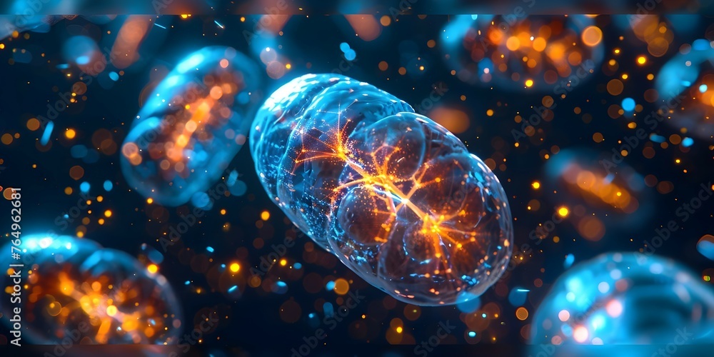
Contrary to many introductory diagrams that render mitochondria as a uniform red or golden mass under standard light microscopes, the true color of mitochondria is far more nuanced. Under high-resolution imaging, including fluoroscopic and electron microscopy, these organelles exhibit a spectrum of hues—from clear and translucent to deep purple, pink, and even greenish-yellow—depending on staining methods, cellular context, and biochemical state.
This variability arises not from inherent pigmentation, but from differences in membrane composition, metabolic activity, and the presence of specific fluorescent cofactors.
The Chromatic Language of Mitochondria: Scientific Imaging and Staining Power
< минMitochondrial coloration is primarily revealed through biochemical staining and advanced imaging techniques that expose molecular details invisible to the naked eye. The most widely recognized visual representation—mitochondria glowing in bright red-orange under fluorescence—stems from supplements like MitoTracker Red CMXRos orToTriton X-100-based tagging, which bind selectively to mitochondrial membranes and report on their membrane potential. These dyes exploit the organelle’s electrochemical gradient, producing vivid fluorescence that paints mitochondria in rich, crimson tones under confocal microscopy.
Yet this color is not intrinsic; it is a learned signature of experimental technique. < min>
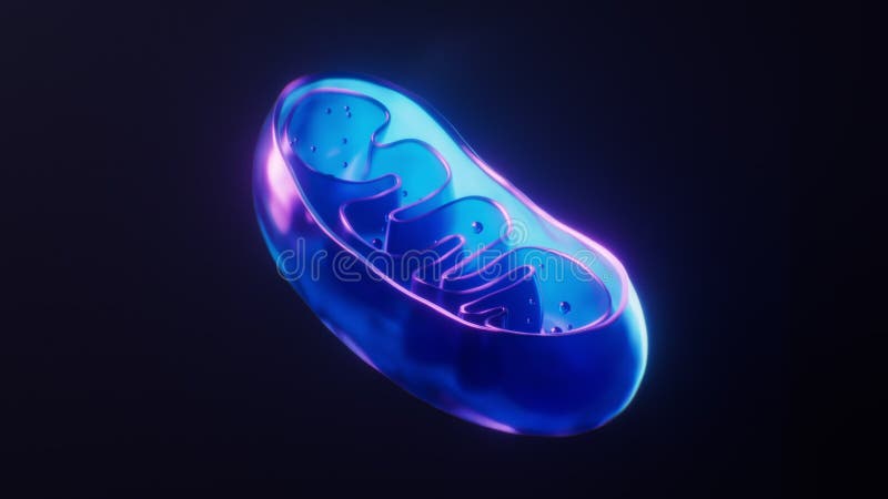
Standard brightfield microscopy typically shows mitochondria as pale, indistinct structures, appearing blue-gray or faintly translucent. This neutral baseline reflects their membranous structure and low lipid content compared to other organelles. However, when scientists use electron microscopy, mitochondria reveal a far more complex architecture—and subtle color shifts.
The inner membrane crests, densely folded for ATP production, sometimes take on darker, speckled contrasts under energy-dispersive X-ray spectroscopy, hinting at localized enzyme concentrations. These dimensional and textural details, though subtle, contribute to a grayscale palette rich with structural information.
< min>More strikingly, mitochondrial color can dynamically shift in response to cellular conditions. During high metabolic demand—such as exercise or neuronal activity—mitochondria swap from a quiet blue-green state into intense red journeys under fluorescence, signaling heightened membrane potential and ATP synthesis.
Conversely, in dysfunction or stress, mitochondrial membranes may lose integrity, causing fluorescence intensity to dim or fragment, revealing disrupted function through color degradation. This chromatic responsiveness makes mitochondrial imaging a powerful diagnostic window into cellular health.
The Role of Chromophores and Electron Transport
Mitochondrial color is deeply tied to the electron transport chain (ETC), where redox-active proteins like cytochrome c and complexes I–IV generate ATP. Flavins and heme groups within these proteins naturally absorb specific wavelengths, contributing to the organelle’s spectral signature.When viewed under advanced fluorescence microscopy, these cofactors produce vivid pink, magenta, and emerald hues, especially in healthy, active mitochondria. The red-shifted emission of cytochrome c (~550 nm) and plastoquinone analogs further enhances the red-orange thermal signature commonly misattributed to “normal” mitochondria. Thus, what appears as "red" often reflects functional status encoded in molecular structure.
Quantifying Color: Tools and Techniques in Modern Cell Biology
< mini>Contemporary cell biologists employ spectrophotometry and image analysis software to decode mitochondrial color with precision. By capturing multi-spectral fluorescence data, researchers can map spectral intensities across mitochondrial networks, transforming color into quantitative metrics. For example, gamma variance analysis of fluorescence intensity across a single mitochondrion reveals metabolic heterogeneity invisible to conventional microscopy.
Machine learning algorithms then classify color patterns to distinguish resting from activated states, or healthy from pathological organelles. This computational color mapping bridges visual science and digital analytics, turning chromatic expression into diagnostic potential.
< mini>Beyond fluorescence, newer label-free imaging methods—such as Raman spectroscopy—detect subtle vibrational signatures tied to mitochondrial lipids and proteins, generating unique spectral fingerprints without dyes. These techniques preserve cellular integrity while revealing subtle color-like contrasts in molecular composition, offering an unbiased glimpse into mitochondrial identity beyond artificial labeling.
The Reality Beneath the Hue: Mitochondria Are Not Actually Pigmented Organelles
Despite recurring metaphors linking mitochondria to colors like “fire,” “blue-green,” or “crimson,” no mitochondria possess pigments.
Their color is not biological but optical—a product of how they scatter and absorb light in imaging environments. This distinction is critical: while the language of color enriches public understanding, it must not obscure the underlying reality—mitochondria are biochemical machines defined by membranes, DNA, and enzymes, not pigments. This nuance underscores a broader principle in science: observation shapes perception, but rigor defines truth.
When Color Tells a Story: Mitochondrial Dysfunction and Color Shifts
< min>Changes in mitochondrial color under the microscope often presage biological dysfunction.
In neurodegenerative diseases like Parkinson’s, mitochondria in dopaminergic neurons lose membrane potential, appearing dimmer and more dispersed under fluorescence. Similarly, in cancer cells with altered metabolism, mitochondrial clustering reveals patchy red fluorescence—an early sign of metabolic reprogramming. Even aging correlates with progressive loss of mitochondrial clarity, as oxidative damage disrupts membrane integrity and fluorescence fidelity.
Tracking these color shifts provides clinicians and researchers with non-invasive indicators of cellular stress.
What This Color Story Means for Science and Medicine
< min>Understanding mitochondrial color—both literal and metaphorical—transforms how scientists interpret cellular health. Far from passive power boxes, mitochondria are active responders, their visual cues reflecting metabolic readiness, redox balance, and structural integrity. In cutting-edge diagnostics, color-based imaging guides precision medicine: identifying mitochondrial defects early, monitoring treatment efficacy, and even predicting disease trajectories.
As fluorescence technologies evolve, mitochondrial color becomes not just a visual phenomenon, but a vital biomarker in the growing landscape of metabolic medicine.
In the end, while the question “What color are mitochondria?” begins with optics, it unveils the intricate language of cellular life—one written in molecular structures, redox reactions, and dynamic visual feedback. Mitochondria are not merely red or purple; they are luminous storytellers, their colors shaped by biology’s deep complexity, molecular choreography, and the ever-changing dance of energy within every living cell.


Related Post
Berniece Baker Miracle Bio Wiki Age Family Husband Daughter and Death
/cdn.vox-cdn.com/uploads/chorus_image/image/36791936/20140106_kkt_bl1_481.0.jpg)
Xavier vs Iowa in College Basketball: A Clash of Rivalries, Lights, and Legacy

Home Depot Marion IL: Your Go-To Hub for Home Improvement Excellence
Jason Colthorp WDIV Bio Wiki Age Height Family Husband Baby Salary And Net Worth