Unlocking the Definition: What Caudal Truly Means in Human Anatomy
Unlocking the Definition: What Caudal Truly Means in Human Anatomy
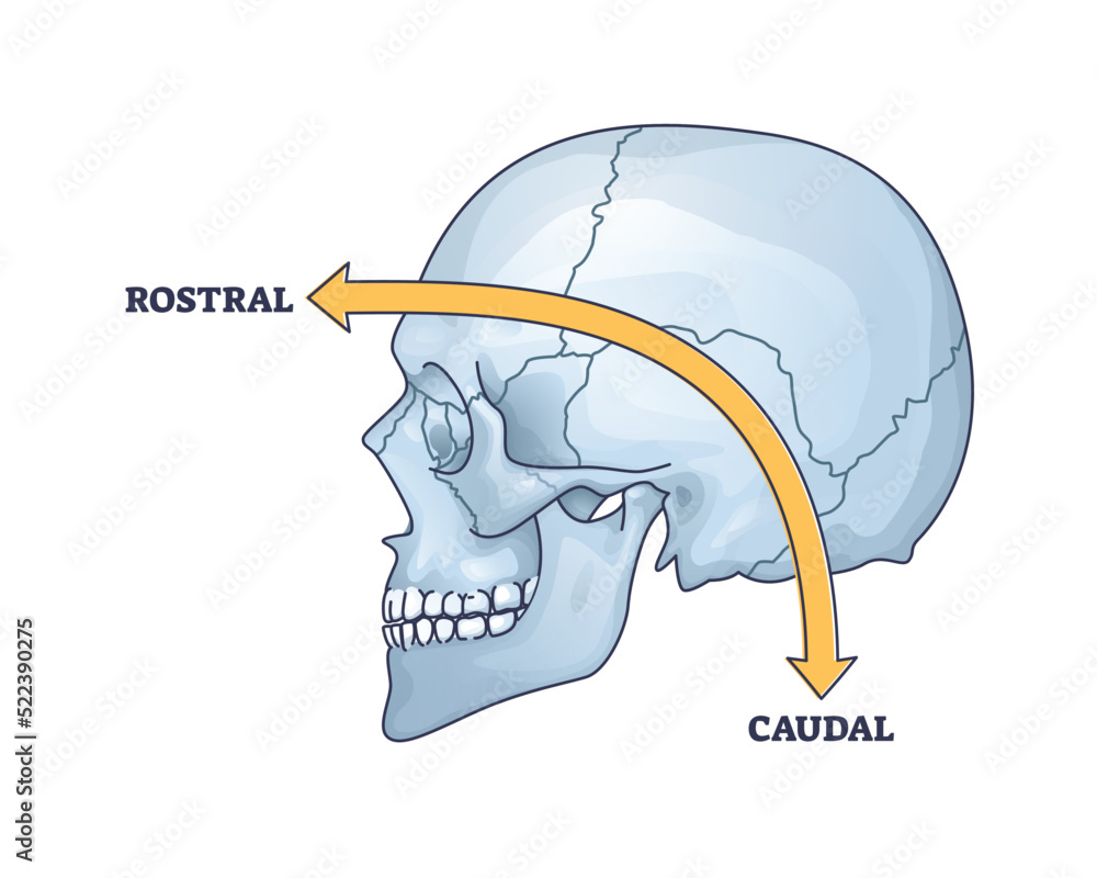
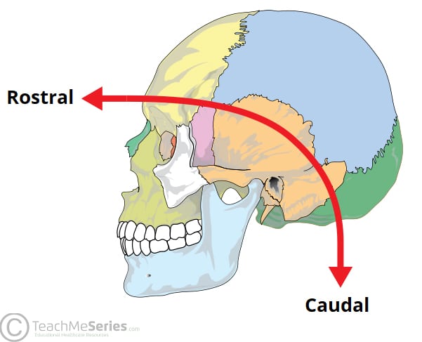
Anatomical terminology often operates like a precise language, enabling scientists, clinicians, and students to communicate with clarity across the complex landscape of the human body. Among the most frequently encountered yet frequently misunderstood terms is “caudal”—a directional indicator rooted in Latin but essential for understanding spatial relationships in anatomical structures. Derived from the Latin *caoudalis*, meaning “lower,” “tail-end,” the term “caudal” designates positions toward the posterior aspect of the body or organs.
Far more than a mere label, caudal provides critical orientation in medical imaging, surgical planning, and neuroanatomy, helping professionals pinpoint locations from upper (cranial) to lower (caudal) with surgical accuracy. Understanding “caudal” requires grounding it within the broader framework of anatomical planes and directional terminology. The human body is conventionally divided into anatomical planes—sagittal, transverse, and frontal—each defining planes of section.
Directional terms then specify positions relative to these planes or key reference points. While “cranial” denotes a position toward the head, the counterpart “caudal” precisely denotes movement or placement toward the tail or lower pole. This duality reflects a bipartite spatial logic that underpins much of anatomical orientation.
The Neuroanatomical Landscape: Caudal in the Spinal Context
Perhaps no domain more rigorously depends on caudal terminology than spinal anatomy. The spinal cord extends from the base of the brain downward through the vertebral column, terminating in the conus medullaris typically around L1–L2 in adults. The segments immediately inferior to this point— clasificándose como *caudal*—form the cauda equina, a bundle of nerve roots jutting from the spinal canal.This region, often likened to a “railway of nerves,” controls sensation and motor function in the legs, pelvis, and lower abdomen. Damage to cauda equina structures—such as from herniated discs, tumors, or spinal trauma—can lead to debilitating conditions like cauda equina syndrome, marked by pain, numbness, and bowel or bladder dysfunction. “A single misplacement in clinical assessment of caudal spinal segments can ignore life-altering pathology,” notes Dr.
Elena Marquez, neuroanatomist at Stanford University. “Precision in labeling caudal levelss is not academic—it’s essential for diagnosis and intervention.” Beyond clinical impact, the caudal designation is integral to developmental anatomy. During embryogenesis, the spinal cord undergoes caudal elongation, a process critical for establishing the neural architecture that governs lower body function.
This developmental gradient reinforces why caudal remains a cornerstone in longitudinal studies tracking neurological maturation and plasticity.
Visualizing the Body: Caudal in Imaging and Surgical Planning
Modern medical imaging modalities—MRI, CT scans, and ultrasound—depend heavily on caudal orientation to interpret structural anatomy in layered detail. Radiologists routinely describe findings using caudal terms to specify lesion location: a mass “caudal to L3” indicates its position relative to the third lumbar vertebra.Similarly, surgical approaches to the spine or sacrum hinge on accurate caudal delineation to minimize nerve damage and ensure precise decompression. “In CT scans, ‘caudal shift’ may signal pathological tilt of spinal segments, altering surgical trajectories,” explains Dr. Raj Patel, orthopedic surgeon at Johns Hopkins.
“Without clear caudal referencing, even minor deviations risk undetected complications.” This level of spatial precision extends beyond the spine. The caudal aspect of organs such as the bladder, rectum, and reproductive structures also follows this logic—each segment extending toward the body’s exit point. For instance, a urological surgeon must distinguish between cranial and caudal ureteral involvement to accurately treat obstructions or diverticula without compromising adjacent nerves.
Caudal in Comparative and Evolutionary Anatomy
The term’s utility spans beyond human medicine into comparative anatomy, where researchers trace evolutionary adaptations across species. Many vertebrates share a fundamental caudal orientation—tail-like posterior appendages essential for locomotion, balance, and sensory input. In fish, the caudal region houses critical swimming muscles; in primates, it evolved toward pendulous structures aiding dexterity and thrashing.Yet in humans, the term “caudal” preserves a lineage of spinal taxonomy, reflecting our unique evolutionary path toward bipedalism and cranial dominance. “Analyzing caudal development in mammals reveals how spinal specialization supports locomotion and posture,” observes Dr. Lin Wei, evolutionary biologist at Cambridge.
“The loss of a pronounced ectocaudal tail in humans is balanced by enhanced caudal spinal control—where flexibility is channeled internally rather than externally.” Such insights reinforce that caudal is not merely a positional descriptor but a window into functional and evolutionary adaptations across the animal kingdom.
In clinical practice, anatomical precision is non-negotiable, and nowhere is this clearer than in the use of “caudal.” Whether localizing a pain source in the lower back, guiding needle placement in minimally invasive procedures, or interpreting imaging data, the term anchors communication in a shared, unambiguous framework. Its Latin root, simple as it may appear, carries profound weight—ensuring that “caudal” is not just defined, but deeply understood.
The Ripple Effect of Caudal Reference in Medical Education
Teaching anatomical terminology carries a pedagogical weight, especially when terms like caudal shape understanding across disciplines.Medical students must internalize cadal positioning not only to pass exams but to translate knowledge into clinical intuition. Simulated lab exercises, 3D modeling, and standardized patient simulations increasingly embed caudal orientation into experiential learning, bridging declarative knowledge with spatial reasoning. “Students who grasp caudal directionality perform better in clinical rotations,” states Dr.
Amina Khalil, leading medical educator at Harvard Medical School. “They visualize procedures more accurately, anticipate complications, and communicate findings with greater clarity—skills that reduce diagnostic errors and improve patient outcomes.” This educational emphasis ensures that caudal remains more than a technical term; it becomes a mental map through which future physicians navigate the body’s intricate architecture.
Ultimately, the meaning of “caudal” transcends dictionary definition—it is a dynamic, functional directional anchor
Related Post
The Gorecenter Conundrum: Navigating the Complex Ethics and Impact of Graphic Content Exposure
A Sprinting Sensation Soars: How Ewa Swoboda Transformed Sprinting from Obscurity to Global Stardom
Paul Finebaum SEC Network Bio Wiki Age Height Family Wife Michigan Show Salary and Net Worth