Mapping the Body’s Core: The 9 Key Regions of the Abdomen
Mapping the Body’s Core: The 9 Key Regions of the Abdomen
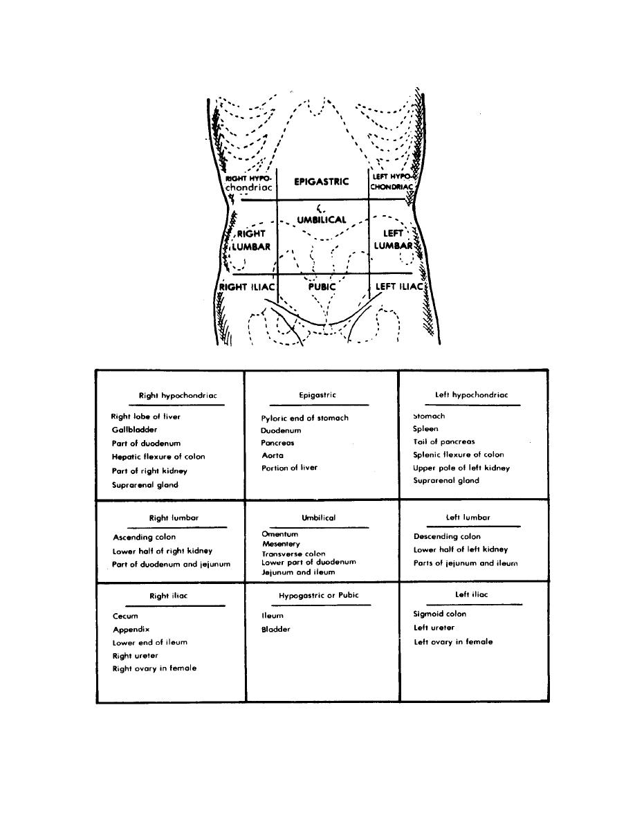
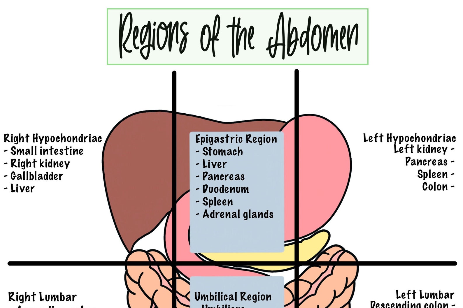
The human abdomen is a dynamic crossroads of vital organs and complex anatomy, serving as a critical interface between respiration, digestion, and mobility. Divided into distinct regions, each section contributes uniquely to physiological function and clinical assessment. Understanding these nine key zones—not merely as labels but as functional landscapes—enhances diagnostics, guides treatment, and deepens appreciation for human anatomy’s precision.
From the protective roof of the upper pole to the mobile tailbone at the base, these abdominal regions form an intricate blueprint essential for both medical professionals and informed laypersons.
1. Upper Right Quadrant (Right Upper Quadrant – RUQ)
The upper right quadrant spans from the costal margin to the ribcage, extending medially into the liver, gallbladder, and right kidney.This region houses the primary site of hepatic and biliary activity, making it pivotal in metabolism and detoxification. Pain here may signal hepatitis, gallstones, or hepatic enlargement. "The RUQ is often the first clue in diagnosing right-sided pathology due to the dense concentration of absorptive and filtering organs," notes a retrospective study in abdominal medicine.
Clinicians frequently palpate this zone to assess liver size and tenderness, framing its diagnostic importance.
2. Upper Left Quadrant (Left Upper Quadrant – LUQ)
Positioned between the sternum and umbilicus, the left upper quadrant contains the spleen, part of the stomach, and portions of the small intestine.It plays a key role in immune function and filtration of aged red blood cells. Stomach pain in this region often correlates with gastritis or gastric ulcers. The spleen’s prominence here means trauma to LUQ can carry serious risks due to vascular density.
As one surgical review states, “The LUQ bridges thoracic and abdominal pressures, making it susceptible to reflux-related discomfort and lymphatic involvement.”
3. Epigastric Region
Centered just below the sternum and spanning to the midclavicular line, the epigastric zone is dominated by the stomach, gastric cardia, and upper portion of the duodenum. This region bears the classic seat of heartburn and indigestion, frequently inflamed in gastroesophageal reflux disease (GERD).Clinically, epigastric pain is a hallmark symptom prompting urgent evaluation for peptic ulcer disease or malignancy. Medical guidelines identify this area as “the waist of the stomach,” underpinning its relevance in diagnosing motility disorders and acid-related conditions.
4.
Right Upper Abdomen (External to RUQ) Beyond the defined upper quadrant lies a broad bowel-related zone encompassing parts of the ascending colon, terminal ileum, and hepatic structures. This transitional segment is sensitive to obstruction, inflammation, or enzymatic irritation. Diarrhea or bloating here often traces to irritable bowel syndrome (IBS) or infectious agents.
This region’s anatomical plasticity makes it vulnerable to visceral hyperalgesia, where minor distension triggers disproportionate pain responses—critical for functional GI disorder assessment.
5. Left Upper Quadrant (Extending Below RUQ)
Below the upper left edge, the LUQ merges with the splenic response zone, housing the distant tail of the pancreas and associated lymphatic passages.While less actively metabolic, this region contributes to lymphatic drainage and immune surveillance. When enlarged, splenic involvement signals systemic conditions such as mononucleosis or chronic hepatitis. Radiologists emphasize that visualizing splenic curvature in the upper left aids early detection of subtle infiltrative diseases.
6. Epigastric Base and Umbilical Junction
This transitional zone spans the epigastric fossa to the umbilicus, integrating frontal and cranial abdominal structures. It serves as a convergence point for nerve modalities, reflex arcs, and visceral sensory feedback.Clinical significance emerges in hernia formation—especially indirect inguinal hernias at this interface. As gastroenterologists clarify, “The epigastric base is where structural integrity and functional feedback intersect, making it indispensable for evaluating abdominal wall integrity.”
7. Right Lower Quadrant (Returning with Terms from LUQ Extension)
Descending from the epigastrium to the inguinal threshold, the right lower quadrant contains the caecum, ileocecal valve, right kidney, and appendix.This region is uniquely susceptible to acute appendicitis and Crohn’s disease. Physicians rely on localized rebound tenderness here to trigger rapid imaging, as delays risk ruptured pathology. The appendix’s confines render this zone a sentinel for inflammatory crises, with clinical guidelines stressing immediate assessment of localized peritoneal signs.
8. Left Lower Quadrant (Adjacent to Bas donna)
In proximity to the spleen and left ureter, the left lower quadrant spans from the body to the pelvis, linking small bowel and colon. It serves as a conduit for diverticulosis, colonic transit issues, and pelvic inflammatory disease.Pain localization here, especially in the sigmoid colon’s distal reach


Related Post

Alfa Romeo 6C Sports Coupe: Where Retro Elegance Meets Modern Craftsmanship
What Cyril Ramaphosas Children Are Up To These Days

Jayson Christopher Tatum Jr: The Rising Star Redefining Basketball’s Next Generation

Globo RJ Ao Vivo Agora No YouTube: Assista Grátis – Tudo Que Você Precisa Saber


